Why Knowing a TF Binding Site Is Only the First Step
Moving from sequence motifs to regulatory mechanisms
Summary
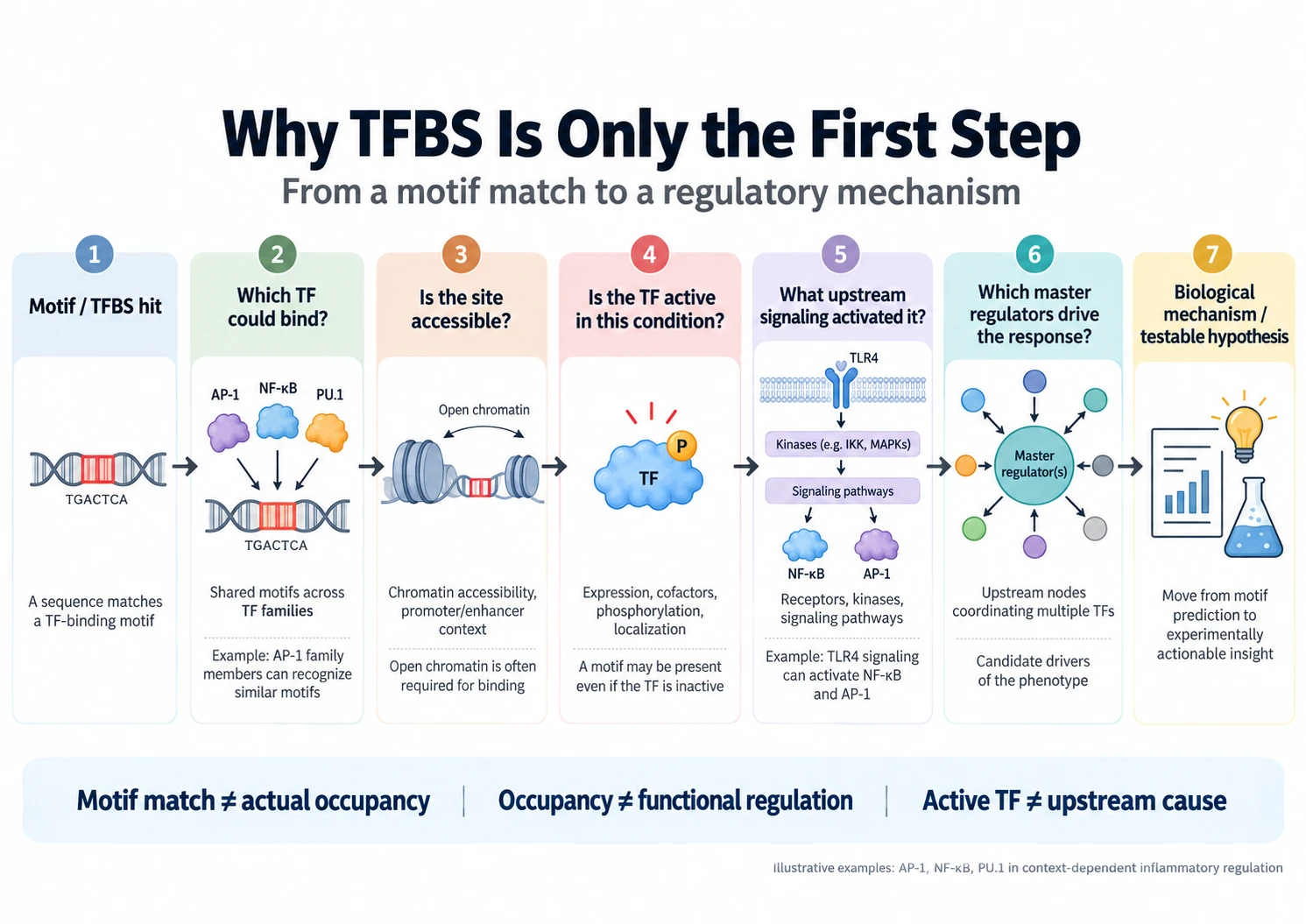
A sequence motif tells us where a transcription factor (TF) could bind. It does not tell us whether the site is accessible in the cell studied, which member of a TF family occupies it, whether that factor is active under the experimental condition, or whether binding changes expression of the nearby gene.
Each of these is a separate question, answered by a separate layer of evidence. Here we focus on the gaps between a motif hit and a regulatory mechanism, and why closing them means following the signal upstream of the TF.
Sequence compatibility is not occupancy
Position weight matrices (PWMs) are the standard tool for locating sequences compatible with TF binding. Scanned across a genome, however, a typical matrix returns far more matches than the factor actually binds. The reason is chromatin. Nucleosome positioning, accessibility, local histone modifications, TF activation by signal transduction as well as the presence of cooperating factors all decide whether a sequence-compatible site can be occupied. DNA shape and flanking sequence add further specificity that a core motif does not capture.
The motif therefore describes a sequence-level potential. The cell state determines whether that potential is used.
A motif often points to a TF family, not an individual factor
Many TF families contain several proteins with closely related DNA-binding domains and almost identical motif preferences. An enriched motif can therefore implicate a TF family without identifying the relevant member, dimer or complex.
AP-1 is a well known example. Jun, Fos, ATF and related proteins recognise essentially the same core element, yet particular heterodimers between individual members regulate different genes and occupy different loci.
Example: AP-1 in macrophages. Fonseca et al. used several AP-1 family members in the ChIP-seq experiments. Factors sharing the same core motif showed overlapping but distinct binding profiles. Moreover, TLR4 stimulation remodelled these profiles. Factor-specific binding was better explained by the local ensemble of collaborating TFs than by the AP-1 motif itself.
“AP-1 motif enriched” is a useful first observation. It is not yet a regulatory explanation.
Even the correct TF may be inactive until an upstream signal activates it
Even the correct TF may be inactive until an upstream signal switches it on. TF activity in many situations is controlled post-translational events: by phosphorylation, acetylation and other modifications as well as by ligand binding, dimerisation, proteolysis, nuclear translocation or co-regulator interactions.
NF-κB is one example. In unstimulated cells, NF-κB dimers are held in the cytoplasm by IκB proteins. Phosphorylation of the IKK complex leads to IκB degradation, which releases NF-κB dimers to enter the nucleus.
A predicted NF-κB binding site is therefore a different biological object in a resting cell and in a cell receiving an inflammatory signal. The sequence is identical; the answer to “is this site occupied?” is not.
Regulatory output is combinatorial and context dependent
Enhancers and promoters usually integrate several TFs rather than acting as one-factor, one-site switches.
In macrophages, inflammatory enhancers combine sites for lineage-determining factors such as PU.1 with sites for stimulus-responsive factors including NF-κB, IRFs and AP-1. Ghisletti et al. showed that this combination tailors inflammatory gene regulation to cell identity. Ostuni et al. then found “latent enhancers”: regions unbound and unmarked before stimulation that become active afterwards.
The regulatory landscape itself therefore changes with the stimulus. The same motif can have very different consequences depending on cell identity, chromatin state and the other TFs bound nearby.
From a motif hit to a mechanistic hypothesis
Each layer answers a different question, and a positive answer at one layer does not settle the next.
| Layer | Question it answers | Typical evidence | What it does not establish |
|---|---|---|---|
| Motif / TFBS | Could a TF bind here? | PWM scanning, conservation | Accessibility, identity of the binding factor |
| Occupancy | Does a TF bind here, in this cell state? | EMSA, ChIP-seq, CUT&RUN, ATAC-seq or DNase footprinting | Whether binding affects expression |
| TF activity & upstream signalling | Which TF or TF combination is functionally active, and what activated these TFs? | Kinase assays, experimental identification of other modifications and protein interactions, pathway analysis, and phosphoproteomics | Why the factor became active and which node is rate-limiting |
| Network driver | Which upstream node could coordinate the response? | Signalling-network reconstruction, then experimental perturbation | Causality, until tested |
Why look upstream of the TFs
When several TFs are implicated in one expression programme, the useful next question is not “which other motifs are present?” It is “what upstream signalling could explain the coordinated activity of these TFs?”
Network reconstruction links receptors, kinases, adaptors and other signalling proteins to the TF layer. Molecules upstream of several affected TFs can then be ranked as candidate control points, referred to as master regulators.
It turns a long list of sequence-level associations into a short list of mechanistic hypotheses that can be tested.
There is also a practical reason. Many TFs lack well-defined small-molecule binding pockets, nuclear receptors being the main exception. Upstream receptors and kinases are often more tractable, so a mechanism that reaches them is also closer to an intervention point.
A concrete biological journey: inflammatory activation of macrophages
Consider a transcriptomic comparison of resting and TLR4-stimulated macrophages. Motif enrichment around the induced genes returns NF-κB, AP-1 and IRF motifs. That result is informative, but it leaves four questions open:
- Which AP-1 family members are responsible?
- Which of the predicted sites are accessible in macrophages?
- Which enhancers existed before stimulation, and which were created by it?
- What signal activates NF-κB, AP-1 and IRFs together?
The mechanistic picture becomes clearer only when the motif layer is connected to macrophage lineage factors such as PU.1 and to TLR4-dependent signalling through adaptor and kinase cascades that activate NF-κB, AP-1 and IRFs. At that point, the analysis begins to explain the observed transcriptional response rather than merely catalogue candidate binding sites.
Why this shaped how we build analysis tools
Regulatory genomics requires multiple analytical layers because each addresses a different part of the mechanism. Motif and TFBS analysis indicate where transcription factors could bind; expression and combinatorial analyses help identify which TFs may be relevant; pathway analysis explores how these factors may be activated; and master-regulator analysis identifies upstream control points where regulatory signals may converge. No single layer is sufficient on its own.
In practice, these steps are often performed in separate tools, making the transitions between them a common point where biological context is lost. This need to move continuously from sequence-level regulation to signalling and network-level mechanisms shaped the design of TRANSFAC Workspace. TRANSFAC supports TFBS and regulatory analysis, while TRANSPATH provides curated signalling and molecular interaction information for upstream and master-regulator analysis. The Genome Enhancer connects these layers so that results from one stage can directly inform the next.
For human disease research, regulatory predictions can be further interpreted using HumanPSD, which links genes and proteins to diseases, biomarkers, drugs, drug targets and clinical-trial information. This makes it possible to examine whether predicted regulators or network components are already associated with a disease mechanism, reported as biomarkers or therapeutic targets, or connected to relevant drugs and clinical evidence.
The aim is therefore not simply to generate more predicted TFBSs or candidate regulators, but to narrow a complex regulatory signal into a smaller set of mechanistically coherent, experimentally testable hypotheses. In this framework, TFBS analysis becomes the starting point for understanding regulatory mechanisms rather than the endpoint.